
Last updated 4mo ago
Log in to add personal notes on this page.
A prenatal ultrasound reveals a single-lobed brain with fused thalami and absent midline structures. The fetus has hypotelorism and a midline cleft lip.
Heterogeneous
- SHH (7q36) - AD, most common single gene
- ZIC2, SIX3, TGIF1
- Also seen in trisomy 13, 18, triploidy
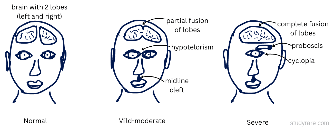
- Spectrum: alobar (most severe) → semilobar → lobar → microform
- Midline facial defects: hypotelorism or cyclopia (one eye), proboscis, midline cleft lip/palate, single central incisor
- Defect of pituitary gland (a midline structure) → loss of pituitary hormones (refer to endocrinology)
- Prenatal ultrasound (single ventricle, fused thalami, absent midline structures) or postnatal MRI defines the structural subtype
- Chromosomal microarray and karyotype to detect trisomy 13, trisomy 18, triploidy, and copy-number variants
- Single-gene or panel sequencing of SHH, ZIC2, SIX3, TGIF1 when chromosomal causes are excluded; assess for maternal diabetes and teratogen exposure
- Parental testing and counseling for autosomal dominant SHH-related disease, which shows wide variable expressivity (an affected parent may have only a single central incisor)
- Varies by severity; hormone replacement for pituitary dysfunction
"The face predicts the brain": Midline facial defects (hypotelorism, cyclopia, midline cleft lip, single central incisor) reflect the severity of the underlying brain malformation. More severe facial findings = more severe brain anomaly.