Mucopolysaccharidosis type 1 (Hurler/Scheie)
On ABGC's listListed on the ABGC Self-Study GuideLog in to starLast updated Nov 10, 2026
A child develops coarse facial features, corneal clouding, hepatosplenomegaly, and developmental delay. Urine shows elevated dermatan and heparan sulfate.
AR; IDUA (alpha-L-iduronidase, a lysosomal enzyme that degrades heparan and dermatan sulfate). Biallelic loss-of-function variants; residual activity sets where a person falls on the spectrum.
Spectrum:
- Hurler (severe): Onset <1 year, coarse features, corneal clouding, severe ID, cardiac disease, death by ~10 years
- Scheie (attenuated): Normal intelligence, later onset, joint stiffness, corneal clouding
- Hurler-Scheie: Intermediate
Alpha-L-iduronidase is a lysosomal glycosidase that removes terminal alpha-L-iduronide residues during the breakdown of heparan sulfate and dermatan sulfate, two glycosaminoglycans (GAGs) of connective tissue. When it fails, both GAGs are stored in lysosomes throughout the body and spill into urine. Storage in the soft tissues of the face and in facial bone gives the coarse features; storage in the oropharynx enlarges tonsils and adenoids and narrows the airway; thickened, stiff valve leaflets cause regurgitation. Beyond storage itself, inflammation, a changed extracellular matrix, and disturbed autophagy and cell signaling contribute.
Genotype predicts phenotype closely:
- Severe: two alleles with no activity, often the common nonsense variants p.Gln70Ter or p.Trp402Ter.
- Attenuated: at least one missense variant in 96% of cases, thought to leave some activity. A few non-missense alleles also leak enzyme, such as c.590-7G>A, which adds a splice site but keeps the normal one.
Management follows the brain. Enzyme replacement (laronidase) does not cross the blood-brain barrier, so it treats the body only; stem cell transplant, best before age 2 years, alters cognitive decline in severe disease.
Part of lysosomal glycosaminoglycan degradation.
Lysosomal degradation of heparan sulfate and dermatan sulfate, by enzymes that each cut a sugar or a sulfate from the end of the chain.
heparan sulfate, dermatan sulfate → lysosome → stepwise removal of end sugars and sulfates → alpha-L-iduronidase (the block; removes terminal iduronide) → further stepwise degradation → free sugars and sulfate
- Accumulates: heparan sulfate and dermatan sulfate, measured as raised urine GAGs; newborn screening measures low enzyme activity in a blood spot.
- Missing: the chain's breakdown products; the disease comes from storage and its effects on the cell.
- Workbook: 7.H. Mucopolysaccharidoses I and II
- Coarse facial features, macrocephaly
- Corneal clouding
- Hepatosplenomegaly
- Dysostosis multiplex (skeletal abnormalities)
- Cardiac valve disease
- On RUSP (some states)
- Newborn screening (some states) measures low alpha-L-iduronidase activity in dried blood spot
- Urine glycosaminoglycans: elevated dermatan sulfate and heparan sulfate
- Confirmatory: deficient alpha-L-iduronidase enzyme activity in leukocytes or fibroblasts, plus IDUA molecular testing
- HSCT (Hurler, if <2 years), enzyme replacement therapy (laronidase)
"Hur1er" = MPS Type 1: the number 1 is hidden in the name. "I did u wrong, I did (iduronidase)": alpha-L-iduronidase degrades dermatan and heparan sulfate.
Hurler has corneal clouding, unlike Hunter (MPS II), who "sees clearly and aims for X." The patient's coarse face makes you want to "hurl." Airway obstruction from glycosaminoglycan deposition in soft tissue of the neck.
Dysostosis multiplex: Malformations of Skeleton aplenty in MPS. All MPS types have bony abnormalities (thick skull, malformed vertebrae, abnormal ribs).
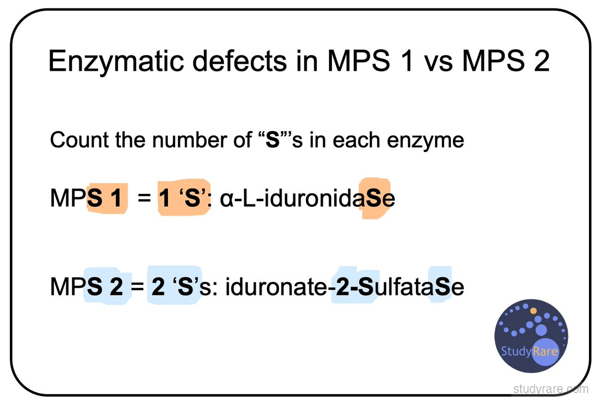
To differentiate MPS 1 from MPS 2, count the number of "S"s in the enzyme name: MPS 1 = 1 "S" (alpha-L-iduronidaSe); MPS 2 = 2 "S"s (iduronate-2-SulfataSe).