Last updated 4mo ago
A 5-year-old African American child presents with severe pain in his arms and legs during a viral illness. He has a history of pneumococcal sepsis at age 2 and his spleen is not palpable.
AR; HBB (beta-globin)
- Glu6Val (HbS)
- Sickle cell disease: HbSS, HbSC, HbS-beta thalassemia
- Vaso-occlusive crises (pain crises)
- Acute chest syndrome
- Stroke (especially children)
- Functional asplenia → infection risk (encapsulated organisms)
- Aplastic crisis (parvovirus B19)
- Sequestration crisis
- Chronic organ damage (kidneys, eyes, bones)
- On RUSP
- Newborn screening detects most cases via the FS pattern (HbF and HbS, no HbA); confirmed by hemoglobin electrophoresis, HPLC, or isoelectric focusing
- HbSS shows predominantly HbS with elevated HbF and no HbA; HbSC and HbS-β-thalassemia give characteristic compound patterns
- Peripheral smear shows sickled cells and Howell-Jolly bodies (functional asplenia); HBB genotyping confirms the diagnosis and supports carrier and prenatal testing
- Hydroxyurea, penicillin prophylaxis, transfusions, HSCT (curative), gene therapy
HbS: p.Glu6Val: Hemoglobin S results from a GAG>GTG transversion at codon 6 of beta-globin (glutamic acid to valine). Hemoglobin C: p.Glu6Lys (same position, different substitution).
Sickle cell management, "Place 1 Sickle into the sorting HHat to get into HuFflePuff": Hydroxyurea increases HbF (fetal hemoglobin), which prevents sickling.
Sickle cell trait: ~60% HbA, ~40% HbS. Carriers are generally asymptomatic but can sickle under extreme conditions.
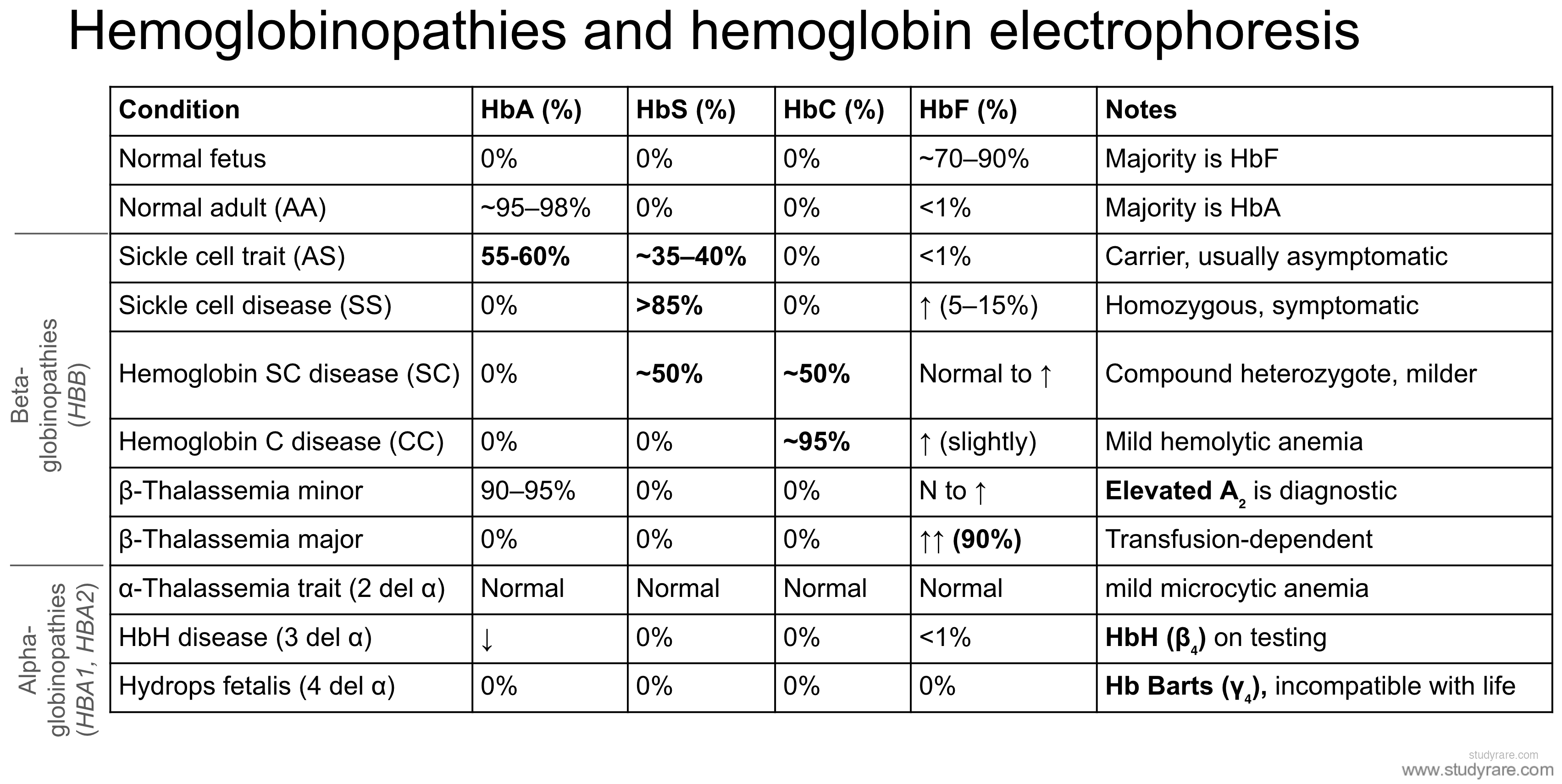
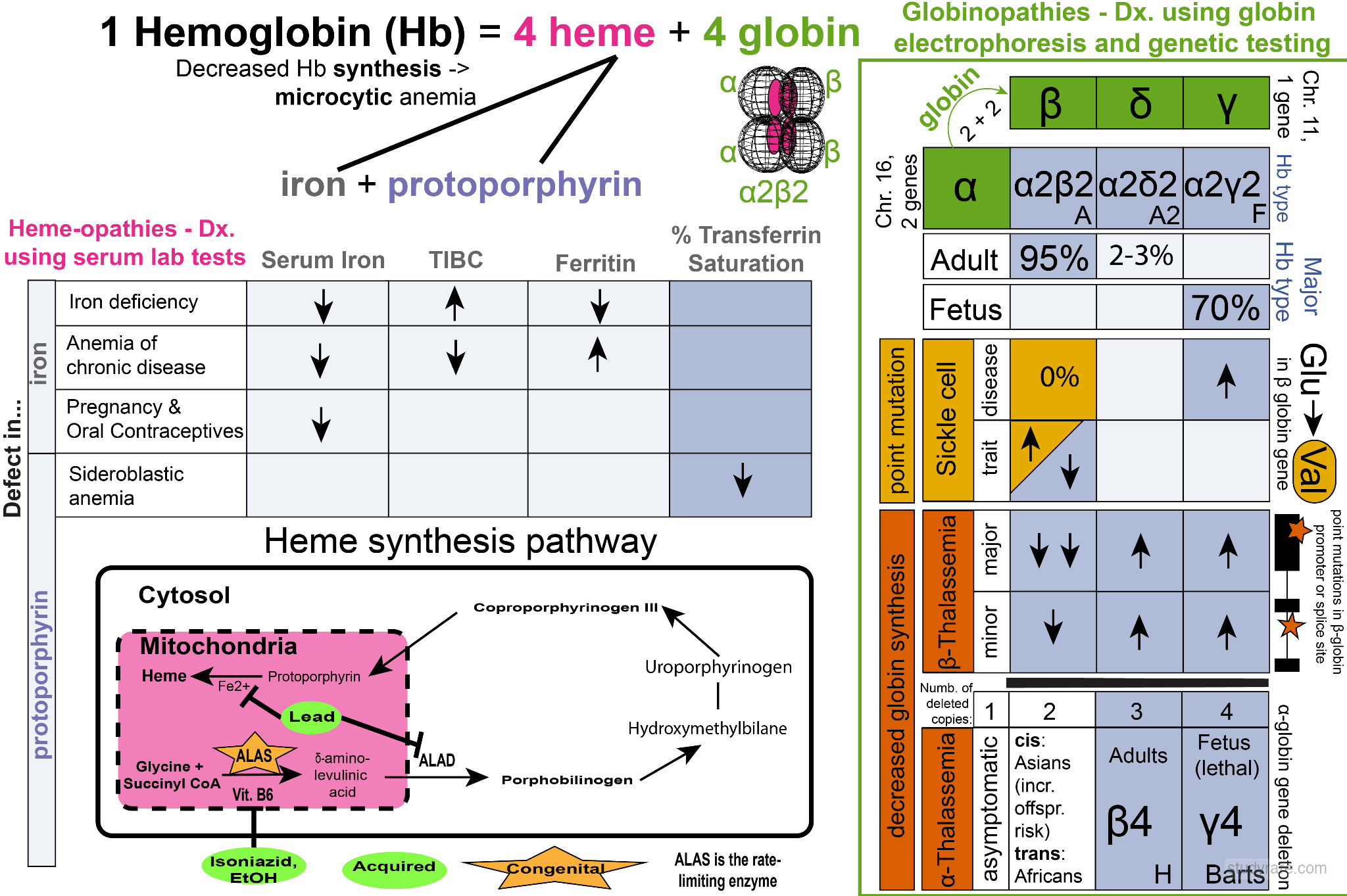
The following overview diagram integrates hemoglobin structure, the heme synthesis pathway, iron studies, and the hemoglobin electrophoresis patterns for sickle cell disease and thalassemias.