Last updated 4mo ago
A 6-month-old Mediterranean infant presents with pallor and failure to thrive. Hemoglobin electrophoresis shows elevated HbF and HbA2. The child requires regular transfusions.
AR; HBB
- β0: No beta-globin production
- β+: Reduced beta-globin production
Types:
- Thalassemia major: Severe anemia, transfusion-dependent
- Thalassemia intermedia: Moderate anemia, variable transfusion needs
- Thalassemia minor (trait): Mild microcytic anemia, usually asymptomatic
- Severe anemia presenting after 6 months (fetal Hb decline)
- Hepatosplenomegaly (extramedullary hematopoiesis)
- Skeletal changes (frontal bossing, maxillary hypertrophy)
- Iron overload (from transfusions and increased absorption)
- Not a named RUSP condition, but often detected incidentally on newborn screening hemoglobin patterns (the RUSP core hemoglobinopathy targets are Hb S,S; Hb S,β-thalassemia; and Hb S,C)
- CBC shows microcytic, hypochromic anemia with normal iron studies (distinguishes from iron deficiency)
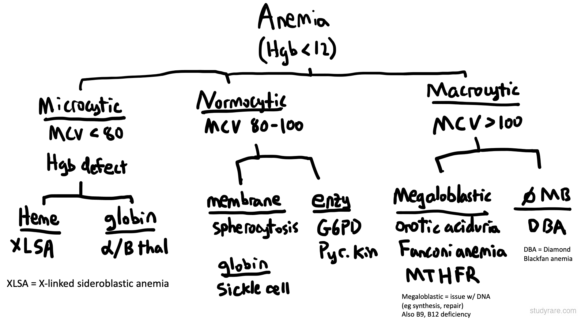
- Hemoglobin electrophoresis or HPLC: elevated HbA2 (>3.5%) is the hallmark of trait, with elevated HbF; major shows markedly reduced or absent HbA
- HBB sequencing confirms the diagnosis and identifies β0 vs β+ variants for prognosis
- Carrier screening and prenatal diagnosis indicated when both partners carry HBB variants
- Regular transfusions, iron chelation
- HSCT
- Gene therapy (betibeglogene autotemcel)
HbA2 is "Up in B diseases": In beta-globin disorders (beta-thalassemia, sickle cell, HbC disease), HbA2 is elevated. Remember: HbA2 is basically HbA (similar name), so it is preferentially made over HbF when beta-globin is mutated.
HbA2 (α2δ2) = 2nd in line to HbA (α2β2). HbA2 Composed of 2 alpha and two delta chains (α2δ2).
When beta-globin is not available to form HbA (because patients have a beta-globinopathy), HbA2 forms
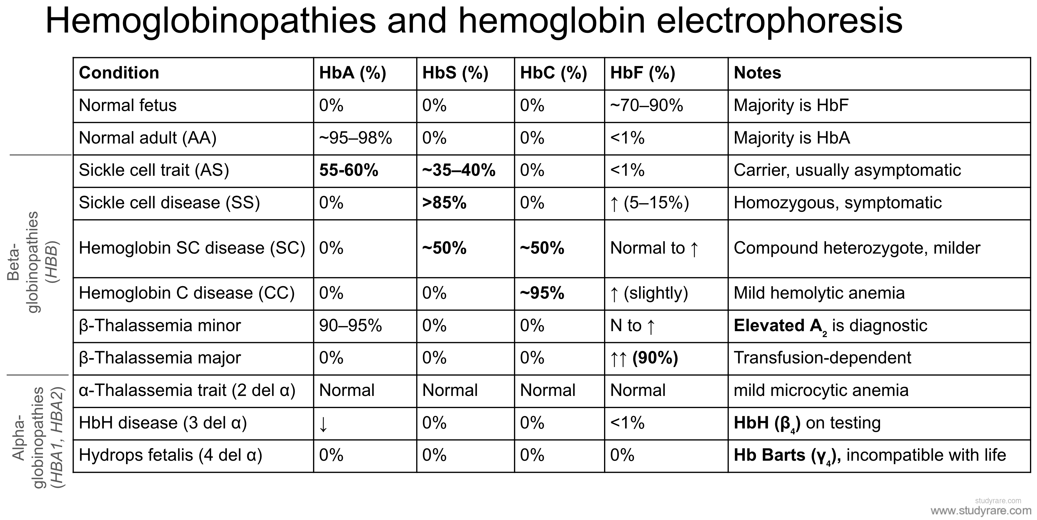
The anemia classification tree organizes anemias by MCV into microcytic, normocytic, and macrocytic categories, with genetic causes highlighted in each group.