
Bone and Connective Tissue Disorders
Overview
This chapter covers disorders affecting the extracellular matrix, skeletal development, and connective tissue integrity. Bone disorders can be organized by whether they affect the extracellular matrix (collagen, hydroxyapatite mineralization) or the cells that build and remodel bone (fibroblasts, osteoclasts, osteoblasts).
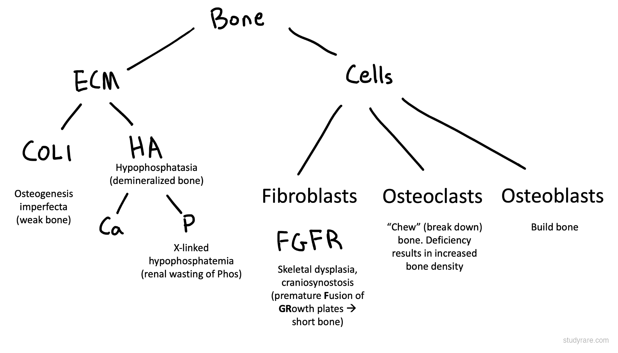
Collagen disorders feature prominently, including the Ehlers-Danlos syndromes (EDS) and osteogenesis imperfecta (OI). Understanding collagen types is essential:
- Type I collagen: bone, skin, tendons (COL1A1/COL1A2 → OI, some EDS)
- Type III collagen: blood vessels, hollow organs (COL3A1 → vascular EDS)
- Type V collagen: skin, classical EDS (COL5A1/COL5A2)
Skeletal dysplasias often involve the FGFR3 pathway (achondroplasia, thanatophoric dysplasia) with activating variants causing impaired endochondral ossification.
Aortopathies (Marfan, Loeys-Dietz) are covered in Chapter 8 (Cardiovascular) but share pathophysiology with connective tissue disorders.
Collagenopathies / Ehlers-Danlos syndromes
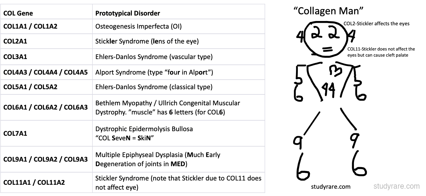
EDS is a group of connective tissue disorders featuring joint hypermobility and skin changes. The 2017 classification includes 13 subtypes, of which a handful account for most cases. Critical point: hEDS is the ONLY subtype without a known gene (clinical diagnosis only). Vascular EDS (COL3A1) is life-threatening due to arterial rupture. The Beighton score assesses joint hypermobility.
Skeletal dysplasias
Skeletal dysplasias are disorders of bone and cartilage growth, often causing short stature and skeletal deformities. The FGFR3 pathway is dominant here: achondroplasia and thanatophoric dysplasia are gain-of-function mutations (more severe = more active). The radiographic features and inheritance patterns distinguish them. Most achondroplasia is de novo, but homozygous achondroplasia is lethal.
Brittle bone disorders
These conditions cause fragile bones prone to fracture. OI involves type I collagen (COL1A1/COL1A2), with severity ranging from mild (type I) to lethal (type II). Blue sclerae, dentinogenesis imperfecta, and hearing loss are associated features. Be aware of the differential diagnosis with non-accidental injury (NAI); genetic testing may be needed to distinguish the two.
Other bone/skeletal
This section covers additional skeletal conditions that don't fit the above categories. HME features benign bone tumors with malignant potential. Fibrodysplasia ossificans progressiva (FOP) is rare but memorable: soft tissue turns to bone, and the big toe malformation is a key diagnostic clue at birth.
Type II collagenopathies
A spectrum of disorders caused by dominant variants in COL2A1, ranging from perinatal lethal (achondrogenesis type II, hypochondrogenesis) through severe (SEDC, Kniest dysplasia) to milder (Stickler syndrome type 1). Most are dominant-negative ("poison-peptide") missense or splice variants in the triple-helical domain. Recognize the shared phenotype (short-trunk short stature, midface hypoplasia, high myopia with retinal detachment risk, sensorineural hearing loss, cleft palate), and remember that Stickler is the milder end of the same spectrum, not a different gene family. Anesthesia precautions (difficult airway from midface hypoplasia + cervical spine instability) apply across the spectrum.
Cohesinopathies
Disorders of the cohesin complex, the multi-subunit protein assembly that holds sister chromatids together during cell division and also plays unrelated roles in transcription regulation. Cornelia de Lange syndrome (NIPBL, SMC1A, SMC3, RAD21, HDAC8) presents with growth retardation, characteristic facies, and limb anomalies, driven by transcriptional dysregulation, not chromosome segregation defects. Roberts syndrome (ESCO2, an acetyltransferase that regulates cohesin) is the dramatic exception: it disrupts sister chromatid cohesion mechanically, producing the diagnostic premature centromere separation (PCS) "railroad track" appearance on standard karyotype plus severe symmetric limb reduction (tetraphocomelia). The PCS finding distinguishes Roberts from CdLS at the bench.
Summary Table
| Disorder | Gene | Inheritance | Cardinal Features |
|---|---|---|---|
| hEDS | Unknown | AD? | Joint hypermobility, pain, no molecular test |
| cEDS | COL5A1/A2 | AD | Skin hyperextensibility, atrophic scars |
| vEDS | COL3A1 | AD | Arterial/bowel rupture, translucent skin |
| kEDS | PLOD1, FKBP14 | AR | Congenital hypotonia, kyphoscoliosis, ocular fragility |
| Stickler | COL2A1, others | AD/AR | Pierre Robin, myopia, retinal detachment |
| Kniest dysplasia | COL2A1 | AD | Short-trunk short stature, knobby joints, dumbbell femora |
| Achondroplasia | FGFR3 | AD | Rhizomelic shortening, macrocephaly |
| Hypochondroplasia | FGFR3 | AD | Mild short-limb short stature, normal facies |
| Thanatophoric | FGFR3 | AD (de novo) | Lethal, severe shortening, narrow chest |
| Diastrophic dysplasia | SLC26A2 | AR | Hitchhiker thumbs, cauliflower ear, cleft palate |
| Cleidocranial dysplasia | RUNX2 | AD | Absent/hypoplastic clavicles, delayed fontanelles, supernumerary teeth |
| Léri-Weill dyschondrosteosis | SHOX | Pseudoautosomal AD | Mesomelic short stature, Madelung deformity |
| OI | COL1A1/A2 | AD (most) | Fractures, blue sclerae, hearing loss |
| Hypophosphatasia | ALPL | AR/AD | LOW alk phos, poor mineralization |
| HME | EXT1, EXT2 | AD | Multiple osteochondromas, chondrosarcoma risk |
| CCA (Beals) | FBN2 | AD | Arachnodactyly, congenital contractures, crumpled ears |
| Roberts syndrome | ESCO2 | AR | Tetraphocomelia, premature centromere separation on karyotype |